ဖိုင်:Brodmann areas 17 18 19.png

အခြားဘာသာစကားများဖြင့် စာမျက်နှာအကြောင်းအရာများကို ပံ့ပိုးမထားပါ။
This is a file from the Wikimedia Commons
ဝီကီပီးဒီးယား မှ

Brodmann_areas_17_18_19.png(၂၅၆ × ၁၉၂ pixels, ဖိုင်အရွယ်အစား - ၄၀ KB, MIME အမျိုးအစား image/png)

အကျဉ်းချုပ်

ဖော်ပြချက်
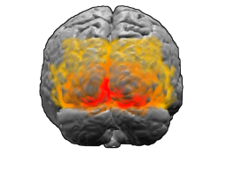
English: Brodmann areas 17, 18 and 19. BA 17 is shown in red. BA 18 is orange. BA 19 is yellow. This is a rear view of the brain. Much of BA 17 is hidden from view on the medial surface (between the hemispheres), on the ventral bank of the calcarine sulcus (check this last point). The brain's surface is extracted from structural en:MRI data (Wellcome Dept. Imaging Neuroscience, UCL, UK). The Brodmann Area data is based on information from the online Talairach demon (an electronic version of Talairach and Tournoux, 1988).
ရက်စွဲ ၁၂ မတ် ၂၀၀၄ (original upload date)
ရင်းမြစ်

These images were created using Blender and Matlab.

. Originally from en.wikipedia; description page is/was here.
စာရေးသူ The original uploader was Washington irving at အင်္ဂလိပ် ဝီကီပီးဒီးယား.

References

  • Talairach, J., and Tournoux, P. Co-Planar Stereotactic Atlas of the Human Brain., New York: Thieme, 1988.

လိုင်စင်သတ်မှတ်ခြင်း

GNU head ဤစာတမ်းကို GNU Free Documentation License, ဗားရှင်း ၁.၂ သို့မဟုတ် Free Software Foundation မှ ဖြန့်ချီထားသော မည်သည့်ယခင်ဗားရှင်းတစ်ခုခုအောက်တွင် Invariant Sections, Front-Cover Texts နှင့် Back-Cover Texts များမပါဝင်ဘဲ ကူးယူခြင်း၊ ဖြန့်ချီခြင်း နှင့်/သို့မဟုတ် ပြင်ဆင်ပြောင်းလဲနိုင်ရန် ခွင့်ပြုသည်။ လိုင်စင်မူပွားကို GNU Free Documentation License ခေါင်းစဉ်အောက်တွင် ထည့်သွင်းထားသည်။
w:my:Creative Commons
မှတ်ယူနိုင်ခြင်း ထပ်တူမျှဝေ
ဤဖိုင်ကို Creative Commons Attribution-Share Alike 3.0 Unported လိုင်စင်ဖြင့် သတ်မှတ်ထားသည်။
သင်သည် လွတ်လပ်စွာ:
  • မျှဝေရန် – ဖန်တီးမှုကို ကူးယူရန်၊ ဖြန့်ဖြူးရန်နှင့် ထုတ်လွှင့်ရန်
  • ပြန်ရောနှောရန် – ဖန်တီးမှုကို ပြင်ဆင်ညှိနှိုင်းရန်
အောက်ပါ အခြေအနေများတွင်:
  • မှတ်ယူနိုင်ခြင်း – ပြောင်းလဲမှုများ ပြုလုပ်ခဲ့ပါက ဖန်တီးသူ သို့မဟုတ် လိုင်စင်ရှိသူမှ သတ်မှတ်သော အမျိုးအစားကို လိုက်နာရမည် (ဆိုလိုသည်မှာ သင့်အား သို့မဟုတ် သင့်ဖန်တီးမှုတွင် အသုံးပြုခြင်းအား ထောက်ခံချက်ပေးသည်ဟု မဆိုလိုပါ)။
  • ထပ်တူမျှဝေ – ဤဖန်တီးမှုအား ပြင်ဆင်ခြင်း၊ ပြောင်းလဲခြင်း သို့မဟုတ် ထပ်ဆင့် ဖန်တီးခြင်း ပြုလုပ်ပါက ရရှိလာသော ဖန်တီးမှုကို ဤဖန်တီးမှုနှင့် သို့မဟုတ် ဆင်တူသော လိုင်စင်ဖြင့်သာ ဖြန့်ချိသင့်သည်။
This licensing tag was added to this file as part of the GFDL licensing update.

မူရင်းတင်ထားသောမှတ်တမ်း

(All user names refer to en.wikipedia)

  • 2004-03-12 22:05 Washington irving 256×192×8 (40769 bytes) Brodmann areas 17, 18 and 19

ပုံစာများ

ဤဖိုင်၏ဆိုလိုရင်းကို စာတစ်ကြောင်းရှင်းလင်းချက်ဖြင့် ထည့်သွင်းရန်

Items portrayed in this file

depicts အင်္ဂလိပ်

media type အင်္ဂလိပ်

image/png

checksum အင်္ဂလိပ်

dfcec62676835d2ab402aea6acdf7ceff9736384

determination method အင်္ဂလိပ်: SHA-1 အင်္ဂလိပ်

data size အင်္ဂလိပ်

၄၀၇၆၉ ဘိုက်

height အင်္ဂလိပ်

၁၉၂ pixel

width အင်္ဂလိပ်

၂၅၆ pixel

ဖိုင်မှတ်တမ်း

ဖိုင်ကို ယင်းနေ့စွဲ အတိုင်း မြင်နိုင်ရန် နေ့စွဲ/အချိန် တစ်ခုခုပေါ်တွင် ကလစ်နှိပ်ပါ။

နေ့စွဲ/အချိန်နမူနာပုံငယ်မှတ်တမ်း ဒိုင်မန်းရှင်းများအသုံးပြုသူမှတ်ချက်
ကာလပေါ် ၀၉:၄၈၊ ၆ ဖေဖော်ဝါရီ ၂၀၀၇ ၀၉:၄၈၊ ၆ ဖေဖော်ဝါရီ ၂၀၀၇ ရက်က မူအတွက် နမူနာပုံငယ်၂၅၆ × ၁၉၂ (၄၀ KB)Siebrand{{Information |Description={{en|Brodmann areas 17, 18 and 19. BA 17 is shown in red. BA 18 is orange. BA 19 is yellow. This is a rear view of the brain. Much of BA 17 is hidden from view on the medial surface (between the hemispheres), on the ventral bank

ဤဖိုင်ကို အသုံးပြုထားသော စာမျက်နှာများမရှိပါ။

ဂလိုဘယ် ဖိုင်သုံးစွဲမှု

အောက်ပါ အခြားဝီကီများတွင် ဤဖိုင်ကို အသုံးပြုထားသည်-

ဤဖိုင်ကို အခြားနေရာများတွင် အသုံးပြုထားမှုများအား ကြည့်ရှုရန်။